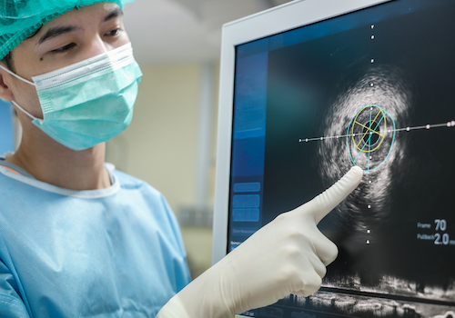
Your heart beats an average of once per second, which makes it one of the hardest working organs in the body. Therefore, seeing it in action is an important part of evaluating and diagnosing disorders of the heart such as heart disease, leaky heart valves and defects in the size and shape of the heart. Various tests help determine what is causing symptoms, help us monitor your heart, and find out if your treatment is working.
Northside Hospital Heart Institute is committed to introducing the most recent technology to provide exceptional patient care and accurate diagnosis of heart disease because we recognize the value of high-quality imaging. For this reason, we utilize sophisticated imaging modalities such as cardiac MRI, cardiac CT, nuclear medicine, and the latest in ultrasound imaging technology.

Comprehensive Medical Imaging of Your Cardiovascular System
At Northside, specially trained radiologists and certified cardiovascular technologists offer a full spectrum of non-invasive diagnostic testing for any heart and vascular need. Using advanced imaging technologies, we provide detailed pictures of your heart, monitor heart function and evaluate how well your veins and arteries are working. Our cardiovascular diagnostic imaging services include:
Diagnostic Cardiac Imaging:
- Cardiac CT: A cardiac CT scan is a non-invasive imaging test that uses X-rays and computer technology to create detailed pictures of the heart and its blood vessels. This scan helps doctors diagnose and assess various heart conditions, plan treatments, and evaluate their effectiveness.
- CT Calcium Scoring: a quick, non-invasive imaging test that uses a CT scan to measure the amount of calcium buildup in the coronary arteries, which can indicate the presence of atherosclerosis (plaque buildup). This score helps assess a person's risk for heart attack or other cardiac events.
- Cardiac MRI: A cardiac MRI is a non-invasive imaging test that uses magnetic fields and radio waves to create detailed pictures of the heart and surrounding blood vessels. It helps doctors assess heart structure, function, and blood flow, aiding in the diagnosis and management of various heart conditions.
- CCTA (Coronary Computed Tomography Angiography: non-invasive imaging test that uses X-rays and computer technology to create detailed 3D pictures of the heart and its blood vessels. This allows doctors to assess the presence, location, and extent of any narrowing or blockages in the coronary arteries, which can indicate coronary artery disease (CAD).
- FFR-CT: is a non-invasive method to assess the severity of coronary artery blockages by evaluating the impact on blood flow. It utilizes CT angiography (CCTA) images and computational fluid dynamics (CFD) to estimate Fractional Flow Reserve (FFR), a measure of blood flow reduction caused by a blockage. This technique allows doctors to determine if a blockage is functionally significant, meaning it's causing a substantial reduction in blood flow, without needing to perform an invasive procedure.
Cardiac Stress Testing:
- Nuclear Stress Test (Pharmacologic & Exercise): a cardiac imaging procedure that uses a radioactive tracer and specialized cameras to visualize blood flow to the heart muscle, both at rest and during stress. It helps doctors identify areas of the heart with reduced blood flow or damage, aiding in the diagnosis of conditions like coronary artery disease. This test can be done by walking on the treadmill or with a medication.
- Cardiac PET/CT: a nuclear medicine imaging test that uses a radioactive tracer and a CT scan to assess blood flow to the heart muscle and identify potential heart problems. It can help diagnose coronary artery disease, assess damage from heart attacks, and evaluate the effectiveness of treatments. The scan combines the structural imaging of CT with the functional information of PET, providing detailed 3D images of the heart and surrounding structures.
- Exercise Stress Test: also known as a cardiac stress test or exercise ECG, assesses how the heart responds to physical exertion. It helps doctors detect heart conditions, particularly coronary artery disease, by monitoring heart rate, blood pressure, and electrical activity (ECG) during exercise.
- Stress echocardiography: A stress echocardiogram is a test that uses ultrasound imaging to assess how well the heart muscle pumps blood while the heart is under stress, typically induced by exercise or medication. It helps doctors diagnose heart conditions, evaluate the severity of valve disease, and assess the heart's response to exercise or medication, especially when a standard electrocardiogram (ECG) might be inconclusive or uninterpretable.
Echocardiography:
- Cardiac Echocardiogram: a non-invasive test that uses ultrasound waves to create images of the heart. It allows doctors to assess the heart's structure, function, and blood flow, helping to diagnose various heart conditions.
- 3D echocardiography and strain imaging: advanced techniques used in echocardiography to assess heart function. 3D echocardiography provides a volumetric view of the heart, while strain imaging measures the deformation of the heart muscle, offering a more sensitive assessment of cardiac function compared to traditional methods. These techniques can be particularly useful in detecting subtle abnormalities and assessing risk in various cardiac conditions.
- Cardio Oncology Echo - cardiac ultrasound that focuses on how cancer treatments affect he heart. This test helps detect heart-related side effects early in patients undergoing chemotherapy, radiation or immunotherapy, allowing for better prevention and management of cardiovascular risks during and after cancer care.
- Transesophageal Echocardiogram (TEE): a medical imaging procedure that uses ultrasound to visualize the heart. It involves inserting a specialized probe (a thin, flexible tube with an ultrasound transducer at the tip) down the esophagus to get a clearer view of the heart than a traditional echocardiogram performed on the chest. TEE is used to diagnose a variety of heart conditions, such as: Heart valve problems, congenital heart defects, Blood clots, Aortic problems, Pericarditis (inflammation of the sac surrounding the heart) and Infective endocarditis (infection of the heart valves or inner lining).
Vascular Ultrasound: a non-invasive imaging test that can help diagnose conditions like blood clots, narrowed or blocked arteries, aneurysms, and varicose veins. It's also used to evaluate blood flow after surgeries like bypasses or to assess the health of dialysis access points.
- Abdominal Aortic Ultrasound: a non-invasive imaging test that uses sound waves to visualize the abdominal aorta, the largest blood vessel in the abdomen. It's primarily used to screen for and diagnose abdominal aortic aneurysms (AAAs), which are bulges in the aorta.
- Carotid Ultrasound: non-invasive imaging test that uses sound waves to visualize the carotid arteries in the neck, assessing blood flow and identifying potential blockages or abnormalities. It helps in detecting carotid artery disease, which can increase the risk of stroke.
- Peripheral Arterial Ultrasound: Evaluates blood flow in the arteries of the arms and legs, identifying blockages, narrowing, or aneurysms.
- Peripheral Venous Ultrasound: Examines the veins in the arms and legs to detect blood clots (deep vein thrombosis or DVT) and assess for venous insufficiency.
Tilt table test: is a diagnostic procedure used to evaluate the cause of syncope (fainting). It helps determine how changes in body position, particularly from lying down to standing, affect blood pressure and heart rate. The test involves tilting the patient on a special table while monitoring vital signs.
Proactive Approach to Heart and Vascular Wellness
At Northside Hospital Heart Institute, we know early detection is the answer to life-long heart and vascular wellness.
Our physicians use the full diagnostic medical imaging capabilities of Northside Hospital Heart Institute to diagnose both routine and complex heart and vascular conditions. We also offer extensive screening tests to assess your risk of cardiovascular diseases. Our screening tests include:
- Coronary artery calcium CT (calcium scoring) to estimate your risk of heart disease
- Exercise stress tests to determine how well your heart responds during exercise while your heart rate is monitored via an electrocardiogram (EKG)
- Lipid panel blood tests to determine your cholesterol and triglyceride levels
- Peripheral artery disease (PAD) ultrasounds to identify the presence of plaque in your limbs
At Northside Hospital Heart Institute, our care team will help you learn how to manage your heart health, such as making simple lifestyle changes to stay healthy – like taking a daily walk and staying more active. While exercise is a crucial component to heart health, diet also plays an important role. We’ll help you learn nutrition tips and create heart-healthy menus that will improve your cardiovascular health.
Cardiovascular imaging and procedures offered at many practice locations and CVDC centers.
Schedule an Appointment
404-845-8200


